

Abstract
This paper focuses on serotonin transporter 5-HTT imaging to investigate major depressive disorder (MDD) and antidepressant occupancy. Such investigations have only recently been possible as a result of major advances in ligand development. The state of the art method is [11C] DASB PET or [11C]-3-amino-4-(2-dimethylaminomethyl-phenylsulfanyl)-benzonitrile) positron emission tomography. [11C]DASB is a breakthrough for brain imaging 5-HTT. Compared with previous radioligands, [11C]DASB offers both high selectivity and a favourable ratio of specific binding relative to free and nonspecific binding. These characteristics contribute to valid, reliable quantitation of the 5-HTT binding potential (BP). The 5-HTT BP can be viewed as an index of 5-HTT density in a medication free state, or unblocked 5-HTT density in a medication-treated state. During major depressive episodes with no other axis I comorbidity, either no difference in regional 5-HTT BP or a trend toward elevated 5-HTT BP is typically found. During major depressive episodes (of MDD) with more severe symptoms of pessimism (dysfunctional attitudes), regional 5-HTT BP is elevated. In subjects with major depressive episodes and comorbid axis I psychiatric illnesses, decreased regional 5-HTT BP is often reported. With selective serotonin reuptake inhibitor (SSRI) treatment at doses that distinguish from placebo in the treatment of major depressive episodes, 5-HTT occupancy is approximately 80%, and there is a strong relation between plasma level and occupancy that is not predictable based on affinity alone. Implications of 5-HTT imaging findings for understanding major depressive disorder and antidepressant treatment will be discussed.
What properties of the serotonin transporter are important for major depressive disorder?
The serotonin transporter 5-hydroxy-tryptamine (5-HTT) is a 630 amino acid long receptor with 12 transmembrane domains. 1,2 The human 5-HTT gene is localized on chromosome 17, centred at 17q11.2.3 Most 5-HTT are located at outer cell membranes, either perisynaptically or along axons.4 In the human brain, the density of 5-HTT varies by region: Superior and inferior raphe nuclei > hypothalamus > thalamus (depending on the nucleus) ~ amygdala > putamen > caudate ~ hippocampus > insular cortex > prefrontal cortex > white matter > cerebellar cortex (except vermis).5–7
The serotonin transporter is coupled to sodium, chlorine and potassium transport.3 However, the physiological role of interest of 5-HTT in major depressive disorder (MDD) and antidepressant treatment is its influence on extracellular serotonin levels. It is clear that many antidepressant drugs that bind to the serotonin transporter raise extracellular serotonin, and 5-HTT knockout mice have elevated extracellular serotonin, confirming the role of the serotonin transporter in modulating extracellular serotonin levels in vivo.8–14
Methods of imaging serotonin transporter in vivo
The following is a critical comparison of all 5-HTT imaging methods that have been applied in humans, with an emphasis on data relevant to humans (See Table 1). Previous comparisons have largely emphasized comparisons in baboons.15 Although this information is valuable during radiotracer development, 16 it does not fully correspond to radioligand performance during human brain imaging because 5-HTT density can vary between animal species,7 and the brain pharmacokinetics of 5-HTT radiotracers can differ between baboons and humans.15,17–21
Comparison of radioligands for imaging of 5-HTT in humans
These methods are used to derive the binding potential (BP). There are different versions of the BP but the one that is typically used is defined as follows: BP = f2 × Bmax/Kd. f2 is a fraction of free and nonspecific radiotracer that interacts with the specific binding compartment. Bmax is receptor density and Kd is the dissociation constant. BP tends to be viewed as an index of Bmax and, in the medication treated condition, it tends to be viewed as an index of receptor density not blocked by medication.
In the medication-treated state, a related measure is the 5-HTT occupancy, which can be defined as 5-HTT occupancy = (5-HTT BP1-5-HTT BP2)/5-HTT BP1 × 100%. 5-HTT BP1 is the BP found in the untreated state and 5-HTT BP2 is the BP found in the treated state.
[123I]2-β-carbomethoxy-3-β-(4-iodophenyl)-tropane (β-CIT) single photon emission tomography (SPECT) was once the only technique developed for measuring the 5-HTT binding potential in humans.37,38,44 This radiotracer has almost equal affinity for the dopamine transporter, compared with the serotonin transporter. 22,23 Because dopamine transporter density is high in the substantia nigra,45 one cannot determine whether any changes in specific binding in the midbrain in an experimental paradigm are due to 5-HTT binding in superior raphe nuclei or dopamine transporter binding in substantia nigra. That there are specific binding sites that are not 5-HTT is consistent with the low 5-HTT occupancy estimates for selective serotonin reuptake inhibitors found with this method,30,31 compared with 5-HTT occupancy estimates with selective 5-HTT binding radiotracers. 34,35 To the best of my knowledge, there are no reliability estimates of binding potential found in the midbrain with this method. Typically, this radiotracer is used for measuring dopamine transporter BP in the striatum in humans.44
The PET radiotracer [11C](+)McN5652 (trans-1,2,3,5,6,10-β-hexahydro-6-[4-(methylthio)phenyl]-pyrrolo-[2,1-a]-isoquioline) shows greater selectivity for the serotonin transporter, compared with other monoamine transporters. It is estimated that this radiotracer has 1 or 2 orders of magnitude greater affinity for the serotonin transporter over the norepinephrine transporter and at least 2 orders of magnitude greater affinity for the serotonin transporter over the dopamine transporter. 24,25 [11C](+)McN5652 has a low ratio of specific binding relative to free and nonspecific binding, which combined with modest reversibility, makes valid and reliable quantitation difficult in regions other than the thalamus, and impossible in the human cortex.18,19,32,39 Applications of this radiotracer in illness and in treatment have mostly focused on the thalamus, using the cerebellum as a reference region with noninvasive models.33,39,46 However, some investigators use arterial sampling to measure 5-HTT BP in other subcortical brain regions to obtain a total distribution volume (an index of total radiotracer binding) and use the cerebellar cortex region to obtain an index of free and nonspecific binding.32
The radiotracer [11C] 3-amino-4-(2-dimethylaminomethyl-phenylsulfanyl)-benzonitrile (DASB) was a major advance because of its selectivity, reversibility, greater specific binding relative to free and nonspecific binding and reliability. 20,21,26,27,34,35,40,43,47 This radiotracer was found to be 3 orders of magnitude more selective for the 5-HTT than for the monoamine transporters and was highly selective for the 5-HTT, compared with several other screened targets.26,27 Moreover, 92% to 95% of the specific binding to 5-HTT is displaceable by 5-HTT binding medications in animal models.26,27 In humans, [11C]DASB has good brain uptake20,40; its ratio of specific binding relative to free and nonspecific binding is good and the latter has low between–subject variability.20,21 Multiple brain regions may be assessed with noninvasive methods,20,21,26,27,34,35,40,43,47 and the reliability of regional 5-HTT BP measures is good.34,35,43,48 The 5-HTT BP measures are low in the cortex, but with standardized region of interest methods, good reliability of 5-HTT BP in the human cortex may be obtained.34,35,43,48 In summary, [11C]DASB PET imaging is the state of the art method in quantifying 5-HTT in humans.
[123I] ADAM (2-((2-((dimethylamino)methyl)-phenyl) thio)-5-iodophenylamine) SPECT is a fourth brain imaging method that has recently been applied to investigate 5-HTT BP in humans. It has a clear advantage of selectivity over [123I] β-CIT SPECT, since most of the specific binding in most brain regions is displaceable in animal models, and it is selective for the 5-HTT over several other binding sites, including other monoamine transporters.28,29 [123I] ADAM has been modelled in baboons but not yet in humans.49 The specific binding relative to free and nonspecific binding in humans is not optimal,41 likely limiting the use of this method to assessing midbrain 5-HTT BP. However, reliability in the midbrain for 5-HTT BP measurement is good.41
[11C]MADAM (11C-N, N-Dimethyl-2-(2-amino-4 methylphenylthio) benzylamine) is a recently developed PET radiotracer that shows excellent selectivity over other monoamine transporters in vitro and good displacability in animal models. 50,51 Time activity curves presented show good reversibility potentially similar to [11C]DASB but appear to have somewhat greater variability, particularly for the raphe.20,52,53 Initial reports of reliability are also promising, although the scatter in repeated-measurement (standard deviation of percent difference in repeated-measure) appears greater than what has been reported for [11C]DASB.34,35,43,48,52,54
What is the optimal method of applying [11C]DASB PET for research protocols?
For selecting regions of interest, my group recommends automated region of interest approaches with visual validation, such as those involving subroutines from linear transformations and/or nonlinear deformations applied in the spatial normalization procedure from statistical parametric mapping. 55,56 Reliability of 5-HTT BP measurement is typically excellent when such applications are applied.35,43,54,57 For subcortical regions, manual drawing upon coregistered MRI also has excellent reliability.34
For a reference region, my group recommends selecting the posterior half of the cerebellar cortex, excluding vermis, excluding white matter and keeping at least one full width half the maximum from the venous sinuses and from occipital cortex. At a distance of one full width half maximum, spillover from the occipital cortex (which possesses specific binding) or venous sinuses is negligible. White matter is excluded because [11C]DASB has different kinetics in this tissue, compared with grey matter. The vermis is excluded because it has [11C]DASB kinetics compatible with significant specific binding. We routinely use these methods.34,35,43,58,59
For selecting models for region of interest methods, we endorse reference tissue approaches.20,21,34,35,43,58,59 By applying a linear regression between 5-HTT density and total distribution volume, we estimate that the reference tissue of posterior cerebellar cortex is composed of 93% free and nonspecific binding and 7% specific binding.6 Knowing that the true BP = distribution volume of specific binding in region of interest divided by the distribution volume of free and nonspecific binding in the cerebellar cortex, the effect of specific binding in the cerebellar cortex is quite subtle. Disease influences of even 50% magnitude on the specific binding in reference tissue translate to 3.5% changes in the distribution volume estimate of free and nonspecific binding, which ultimately results in a 3.5% bias for between–group comparisons. For occupancy studies, the nature of the occupancy equation is such that the bias from a 7% underestimate during untreated conditions is translated into a lesser bias in the overall occupancy measure (less than 2%). For example, if the striatal 5-HTT BP has a true value of 1 in the untreated condition and 0.2 in the SSRI-treated condition, the true 5-HTT occupancy is ([1–0.2]/1) = 0.8 or 80% (5-HTT occupancy = (5-HTT BP1–5-HTT BP2)/5-HTT BP1 × 100%). Taking into account the slight specific binding of reference tissue, the measured striatal 5-HTT BP, respectively, would be 0.93 in the untreated condition and 0.197 in the SSRI-treated condition (most of the 7% specific binding in reference tissue is blocked during treatment), leading to a measured 5-HTT occupancy of ([0.93–0.1972]/0.93) = 0.79 or 79%.
For [11C]DASB PET, arterial methods offer no advantage for identifying subcompartments of free and nonspecific binding, because [11C]DASB kinetics fit a single tissue compartment model in all regions.21,60 Arterial methods do permit measurement of total distribution volume in the cerebellum, but this value is assumed to represent free and nonspecific binding, so as to quantitate binding potential measures in other regions. Thus, when arterial sampling is done, a very similar set of assumptions as compared with reference tissue models are applied.
Among the reference tissue methods, the noninvasive logan, 61 simplified reference tissue model 2 and multilinear reference tissue model 221 have excellent reliability.34,35,43,54,58,59 The latter two also avoid underestimating.21 The Logan has some underestimate but correlates highly with the ratio of the distribution volume in regions with specific binding to the distribution volume in the cerebellum.35 Moreover, with the Logan, the coefficient of variation is very low, and it has less assumptions (i.e., it does not require the single tissue compartment model).61,62 For region of interest measurement in disease processes, we favour all 3 methods, but for drug-treated conditions, we prefer the Logan (to avoid requiring the single tissue compartment assumption across different levels of 5-HTT occupancy).62
What is the key evidence for low extracellular serotonin in untreated MDD?
Direct evidence that serotonin is low in MDD is unavailable for 2 main reasons: Brain serotonin cannot be directly measured in vivo and it is likely, based on animal simulations of postmortem delay, that serotonin levels are unstable, even within 24 hours of death.63 Moreover, postmortem investigations of serotonin levels (previously listed by Mann64) have not sampled medication-free subjects with MDD in the midst of a major depressive episode (MDE).
Therefore, arguments that extracellular serotonin in the brain is likely to be low during MDE are based on the reversal of symptoms after serotonin-raising antidepressant drugs,65–68 lowering of mood during paradigms that lower brain serotonin,69–77 and changes in indices of serotonin 2 receptor density in suicide and MDD.78–90
This latter argument can be further clarified. An important property of 5-HT2 receptors is that 5-HT2 receptor density has an inverse relation to extracellular serotonin levels, such that the density of 5-HT2 receptors in the cortex increases after chronic serotonin depletion and decreases after chronically raising extracellular serotonin.91–94 Therefore, investigations of indices of 5-HT2 density would be expected to report elevations in the midst of MDE. Postmortem investigations sometimes report elevated 5-HT2 density in the prefrontal cortex of suicide victims,78–88 and several of the investigations that found elevated prefrontal 5-HT2 receptor density investigated subjects with MDD.81,84 Such studies could be considered supportive of low extracellular serotonin in the prefrontal cortex of subjects with MDD.
Studies of 5-HT2 receptors in the brain cortex usually measure 5-HT2A receptors because ligand binding to 5-HT2C receptors in the cortex is extremely low95,96 and mRNA of 5-HT2B receptors is extremely low in the cortex.97
At first review, there appears to be a contradiction between the results of postmortem and brain imaging studies of cortex 5-HT2 receptors in MDD. Most of the imaging studies listed in Table 2 report a regional decrease in 5-HT2 BP. The discrepancy can be resolved partly by the observation that most of these studies sampled subjects recently treated with serotonin-raising antidepressant drugs.
Imaging studies of 5-HT2A receptors in major depressive disorder
Upon further review of Table 2, the studies sampling subjects who recently had antidepressant treatment tend to report decreased regional 5-HT2 BP, whereas the 2 studies not sampling subjects who recently had antidepressant treatment find no difference between subjects with depression and healthy subjects.101,102 The study by Meyer and colleagues applied [18F]setoperone PET. [18F]setoperone is a good radioligand for imaging 5-HT2A receptors because of its specific binding in the cortex, reversibility and favourable ratio of specific binding to free and nonspecific binding106–110 (see Table 3 for properties of [18F]setoperone). It is also insensitive to acute paroxetine-induced changes in extracellular 5-HT in humans.118 The study by Meyer and colleagues sampled medication-free (> 6 mo), early-onset depression subjects with no comorbid psychiatric illnesses and found no difference in the prefrontal cortex 5-HT2 BP, compared with healthy control subjects.101 An investigation using [18F] altanserin PET in older subjects with depression who were medication free similarly found no difference in 5-HT2 BP between patients and healthy control subjects.102 After considering medication-free status, there was still a lesser discrepancy, such that 5-HT2 density was often elevated in the prefrontal cortex in postmortem studies of suicide victims, yet prefrontal 5-HT2 BP was not changed in medication-free subjects with depression.
Properties of [18F] Setoperone
To resolve this lesser discrepancy, a more complicated model of low-cortex serotonin during MDEs was hypothesized. This hypothesis was that extracellular serotonin loss is heterogenous during depressive episodes and that the loss is most severe in people with a greater severity of particular symptoms.
The symptom chosen in this hypothesis was elevated pessimism (dysfunctional attitudes) observed during MDEs. (There is a modest level of dysfunctional attitudes that increase during depressive episodes.) The rationale for choosing elevated dysfunctional attitudes is that raising extracellular serotonin after administering intravenous d-fenfluramine is associated with a strong shift in dysfunctional attitudes toward optimism 1 hour later in healthy individuals.89 This suggests that, among the many roles of serotonin, one of them is to modulate dysfunctional attitudes in humans.
Dysfunctional attitudes can be measured with the dysfunctional attitudes scale (DAS), a measure sensitive for detecting negative thinking in the midst of depressive episodes119,120 that has good internal consistency (Cronbach’s α = 0.85 to 0.87)121,122 and high test-retest reliability.122,123
If the hypothesis were true that extracellular serotonin in the cortex is variably reduced in the midst of depressive episodes, with the largest reductions in people with the most severe dysfunctional attitudes (pessimism), one would expect the highest prefrontal 5-HT2 BP in subjects with the most severe dysfunctional attitudes (pessimism). This is based on the finding that 5-HT2 receptor density increases after long-term serotonin depletion.92,93 In support of the hypothesis, a strong correlation was observed between severity of dysfunctional attitudes (pessimism) and elevation in cortex 5-HT2 BP.89 Moreover, cortex 5-HT2 BP was significantly elevated in subjects with severe depression with severe pessimism. For example, in the prefrontal cortex region centered on Brodman’s area 9, 5-HT2 BP was elevated 29% in depression subjects with dysfunctional attitude scores higher than the median for the group (Fig. 1). Thus the 2 parts of the study combined argue that extracellular serotonin is low in depression subjects who have a greater severity of pessimism.89
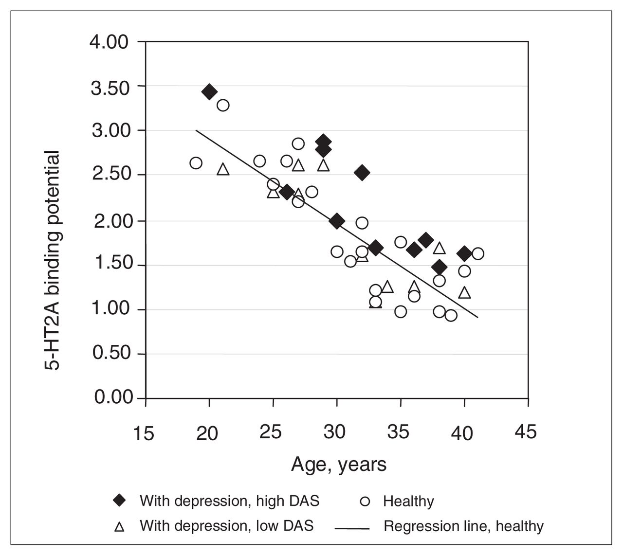
5-HT2A receptor binding potential in averaged bilateral middle frontal gyrus (Brodmann’s area 9) is plotted against age to show the relation between depressed and healthy subjects. The 22 depressed patients were divided into high and low dysfunctional attitudes scale (DAS) groups, depending on whether their DAS scores were above or below the median DAS score for depression patients. This median score was 166. Patients with high DAS scores had significantly higher 5-HT2A receptor binding potential, compared with healthy subjects (ANCOVA [age covariate], diagnosis, F1,19 = 11, p = 0.003). Reprinted with permission from the American Journal of Psychiatry.
This investigation89 is the first imaging study to show consistency with postmortem investigations reporting increased 5-HT2 receptor density in the prefrontal cortex: The dysfunctional attitudes scale is well correlated with hopelessness, as measured with the Beck Hopelessness Scale.124–127 Given that hopelessness is a risk factor for suicide,128,129 it is plausible that investigations of suicide victims reporting increased 5-HT2 BP sampled depression subjects with a greater severity of pessimism. A recent report has replicated the relation between cortex 5-HT2A BP and severity of dysfunctional attitudes in recovered depression patients.130
Serotonin receptor binding that relates to extracellular serotonin in subcortical regions is also important. An interesting postmortem study by Stockmeier and colleagues90 found elevated [3H]OH-DPAT binding in the dorsal raphe nucleus of subjects in the midst of depressive episodes. Replication of this finding has been reported in suicide victims.131 [3H]OH-DPAT, being a 5-HT1A agonist, would be expected to have elevated binding when serotonin is low.132 In this interpretation, the result would support a hypothesis of lower extracellular serotonin in the dorsal raphe region (although this finding could also be viewed as a mechanism that enhances inhibition of the dorsal raphe by nearby extracellular serotonin90).
Possible disease models and the serotonin transporter in MDD
If extracellular serotonin is low during MDEs, with greater severity of dysfunctional attitudes, then it is logical to consider serotonin transporter function in the removal of extracellular monoamines. There are at least 4 plausible ways to understand how 5-HTT BP, an index of 5-HTT density and affinity, could be altered in a disease that lowers brain serotonin, described below. Prior to investigations of [11C]DASB imaging during depressive episodes, model 1 and model 3 were the most plausible.
Model 1
The first possible model is a lesion model. If serotonin nerve terminals were destroyed in MDD, then one could expect that there would be less release of serotonin. This model is observed in neurotoxin exposure133 and late in Parkinson’s disease.134,135 If there were less nerve terminals in MDD, then one would expect a reduction in regional 5-HTT BP.
Model 2
A second model to consider would be whether a lowering of extracellular serotonin (via a process unrelated to 5-HTT sites) would have a secondary effect on 5-HTT density. Acute reductions in serotonin have repeatedly shown reductions in 5-HTT mRNA.136–138 However, longer-term reductions or elevations in serotonin typically show no influence on regional 5-HTT density.139–141 The is not comparable to other monoamine transporters. For example, for the dopamine transporter, the evidence is much stronger to support a relation between long-term reductions in extracellular dopamine and a lowering of striatal dopamine transporter density.142–145 This second model seems unlikely.
Model 3
A third model is increased clearance of serotonin via greater density of 5-HTT. There is an inverse relation between functioning 5-HTT and extracellular serotonin. For example, it has been demonstrated through antidepressant occupancy and 5-HTT knockout models that less functioning 5-HTT are associated with greater extracellular serotonin.9–14,146 It is possible that, under conditions of greater 5-HTT density, greater extracellular serotonin loss may occur. With this model, greater 5-HTT BP would be associated with more severe serotonin loss.
Model 4
A fourth model to consider is endogenous displacement. Endogenous displacement refers to the property, found in a minority of PET radiotracers under physiological conditions, to have increased binding potential measures after a reduction in endogenous neurotransmitter.147 The name originates from the initial explanation for this phenomenon, that the neurotransmitter itself prevented access of the radiotracer to receptors. For [11C]DASB, endogenous displacement may occur with large magnitude changes in extracellular 5-HT but would not be expected to occur with extracellular 5-HT changes that are physiologically relevant for humans. In an animal study with [11C]DASB, after raising extracellular serotonin with an intraperitoneal injection of 10 mg/kg of tranylcypromine, a MAO-A and MAO-B inhibitor, a reduction in 5-HTT BP was observed.148 Similar work has been replicated with similarly substantial doses of tranylcypromine.149 Notably, the rise in extracellular serotonin with high doses of tranylcypromine is enormous, with a several hundred to thousand percent rise being typical.150–152 Humans cannot tolerate 1/10 this dose of tranylcypromine, even with lengthy titrations and oral administration. Thus this magnitude of serotonin change may exceed what is physiologically relevant in humans. In 14 humans, we examined the effect of tryptophan depletion upon 5-HTT and found no effect, demonstrating that endogenous serotonin occupancy is unlikely to appreciably influence [11C]DASB43 under physiologically tolerable conditions. Talbot and colleagues reported similar results in 8 humans.153 Hence, the fourth model is unlikely to apply to PET imaging studies with [11C]DASB in humans.
Studies of the serotonin transporter in MDD
There are only 2 postmortem investigations of 5-HTT density in subjects with recent symptoms of depressive episodes. In these 2 studies, no changes in 5-HTT density were found in either the dorsal raphe or the locus coeruleus.154,155 Other postmortem studies of 5-HTT density sampled subjects with a history of a depressive episode (not necessarily recent) and typically focused on prefrontal cortex regions. These studies tend to report either decreased 5-HTT density156–159 or no difference in 5-HTT density.160–165 In several of these studies, subjects were medication free, based on clinical history and toxicological screening.158,160,161 For many of these investigations, average postmortem delays were less than a day.154–156,158,159,162
Most of these studies were recently reviewed in detail by Stockmeier and colleagues.88 However, most postmortem studies are not representative of a depressive episode, since only 2 studies of 5-HTT density sampled subjects who recently had symptoms.154,155 Further, the postmortem studies are not completely selective for MDD because all include comorbid axis I psychiatric illnesses156–162 and some also sample bipolar disorder. 158,160,165 A third key issue with sampling is that all studies include both early-and late-onset MDD.156–162
The first imaging study of 5-HTT in vivo used [123I]β-CIT SPECT to measure specific binding of [123I]β-CIT in the hypothalamus-midbrain region in depressed patients and healthy control subjects.166 See Table 4 for a descriptive list of imaging studies of the 5-HTT. A reduction in the binding potential was found. Interestingly, subjects with major depression were subdivided based on recency of medication use, and this did not affect the results. The following methodological issue makes this finding difficult to interpret: Because [123I]β-CIT has similar affinity for both 5-HTT and DAT transporters166 and because both the raphe and substantia nigra are within this region sampled, it is not clear whether specific binding of β-CIT reflects 5-HTT binding or DAT binding. The DAT downregulates in striatum after long-term dopamine depletion.142–145 If DAT in substantia nigra downregulate similarly and if there is a monoamine lowering process in the midbrain during depressive episodes, it would be expected that an index of specific binding (to serotonin and dopamine transporters) would be lower.
Imaging investigations of the serotonin transporter in untreated major depressive episodes
The next study of 5-HTT applied [11C](+)McN5652 PET to measure thalamus 5-HTT BP in 7 subjects with MDD.46 The data from these subjects were added to data from 6 subjects who had bipolar disorder. Ichimiya found that, in subjects with either MDD or bipolar disorder, 5-HTT BP in the thalamus is elevated.46 A strength of the study was that subjects were medication free, and a more selective radioligand was used. A disadvantage of the study is that it may be incorrect to assume that unipolar MDEs and bipolar MDEs have a common serotonin transporter abnormality. This study did not investigate 5-HTT BP during a current MDE because only 5 subjects with a current MDE and MDD were enrolled in the study.46
The next study was the first application of [11C]DASB PET imaging to MDD. It sampled 20 subjects with MDE and 20 healthy controls.170 This sample had a number of advantages because it was reasonably large, subjects were medication free for at least 3 months, and they had no other comorbid axis I illnesses, were nonsmoking, and had early onset depression. As a result of the technical advantages of [11C]DASB, 5-HTT BP could be reliably measured in multiple brain regions, including cortex. There was no evidence to support a hypothesis of a difference in 5-HTT BP as no difference in regional 5-HTT BP was found between MDE and healthy subjects in any brain region.170
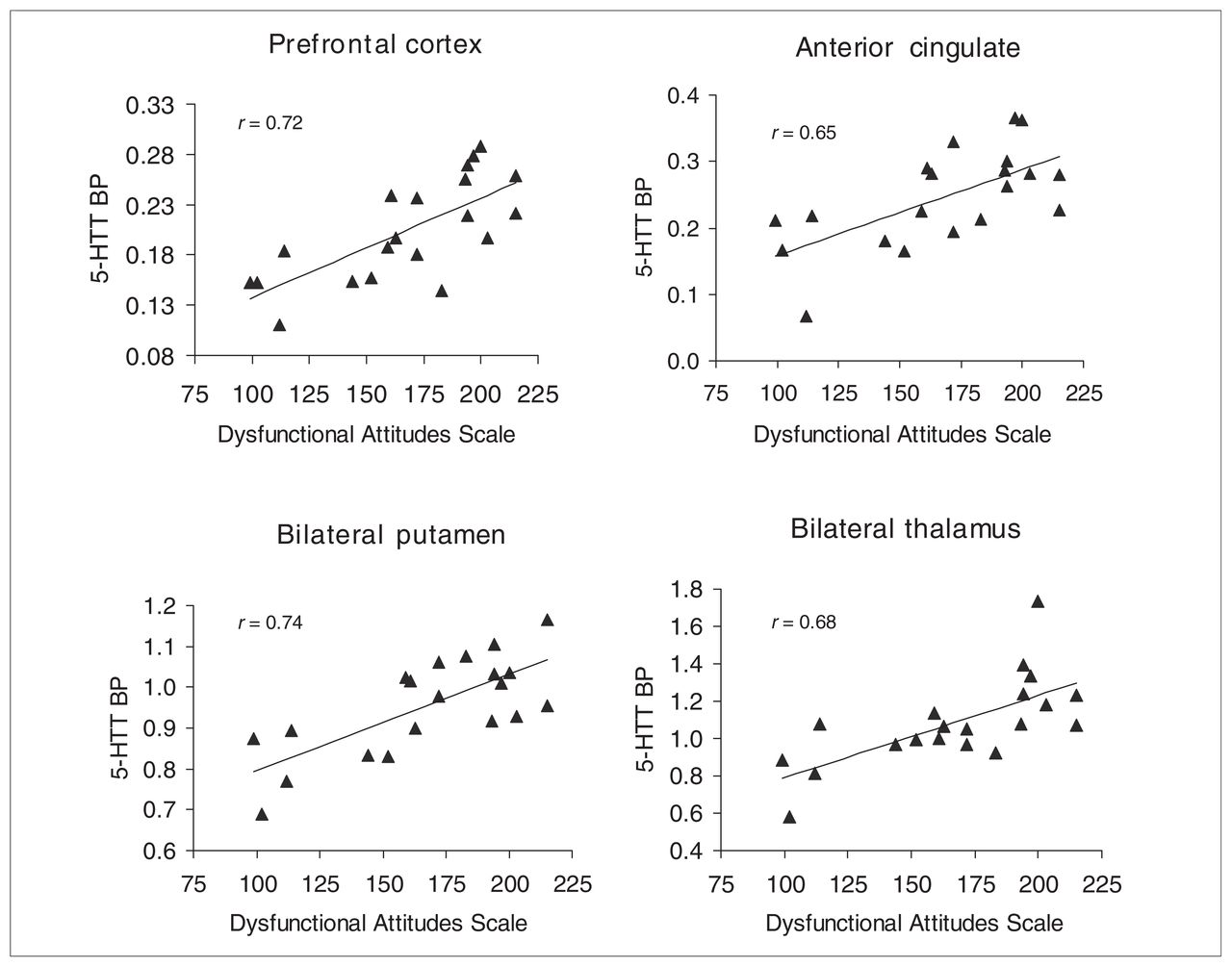
However, in this same study of [11C]DASB PET,170 there was highly significant support for the hypothesis that greater regional 5-HTT BP would occur during MDE with severe, pessimistic dysfunctional attitudes. This hypothesis was based on the interpretation of 3 findings which argue that extracellular serotonin is lowest during MDE with severe, pessimistic, dysfunctional attitudes: The first finding is the acute shift toward optimism in humans after raising extracellular serotonin with d-fenfluramine which argues for a role of serotonin in modulating pessimism/optimism in humans.89 The second finding is that cortex 5-HT2 BP is greater during MDE with severe pessimism.89 This can occur when extracellular serotonin is low, according to the third set of findings: 5-HT2 receptor density increases when extracellular serotonin is chronically lowered92,93 (for more detail, see Key Evidence For Low Extracellular Serotonin in Untreated Major Depressive Disorder in this article). The subgroup of MDE subjects with severely pessimistic dysfunctional attitudes had significantly higher 5-HTT BP, compared with healthy subjects, in brain regions sampling serotonin nerve terminals (prefrontal cortex, anterior cingulate, thalamus, bilateral caudate, bilateral putamen) (see Fig. 2). On average, 5-HTT BP was 21% greater in these regions in MDE subjects with severely pessimistic dysfunctional attitudes (see Fig. 3). Moreover, within the MDE group, greater 5-HTT BP was strongly associated with more negativistic dysfunctional attitudes in the same brain regions. The interpretation was that serotonin transporters have an important role in influencing extracellular serotonin during MDEs: Greater regional 5-HTT BP can provide greater vulnerability to low extracellular 5-HT and symptoms of extremely negativistic dysfunctional attitudes.170

Correlations between dysfunctional attitudes and serotonin transporter binding potential (5-HTT BP) in some of the larger regions in depression subjects. Highly significant correlations were found: prefrontal cortex (p = 0.0004), anterior cingulate (p = 0.002), bilateral putamen (p = 0.0002), bilateral thalamus (p = 0.001). Reprinted with permission from the Archives of General Psychiatry.
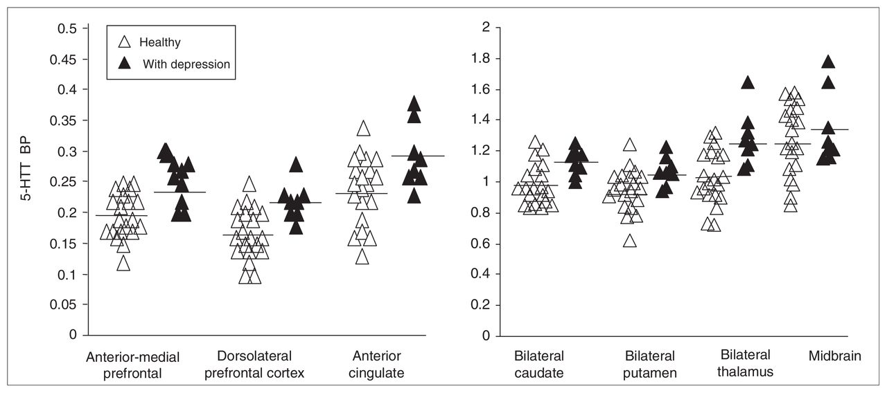
Comparison of regional 5-HTT BP between 8 subjects with depression with severely negativistic dysfunctional attitudes (greater than 190) and 20 healthy subjects. For regions primarily sampling serotonergic nerve terminals (prefrontal cortex, anterior cingulate, caudate, putamen, thalamus) the 5-HTT BP was significantly greater in the depressed group (F1,26 = 5.6 to 12.2, p = 0.03 to 0.002). The midbrain 5-HTT BP was not significantly different (F1,26 = 0.5, p = 0.5). Reprinted with permission from the Archives of General Psychiatry.
It is premature to conclude upon the etiology of an elevation in 5-HTT BP in the subset of MDE subjects with more severe dysfunctional attitudes, because one could consider both genetic171 and environmental influences.172 The simplest explanation is that the subgroup with greater pessimism happened to inherit a greater 5-HTT density. Under this explanation, it would be expected that inheriting a greater 5-HTT could increase the risk of acquiring a MDD with more severe pessimism during MDEs.
The relation between genotypes such as the 5-HTTLPR and/or 5-HTT LPR (LA/LG) and brain 5-HTT density or binding potential is an area of ongoing study.59,158,173–178 Some investigators interpret the genotype associated with greater 5-HTT synthesis in cell lines as being reflective of 5-HTT density in the brain. The genotype associated with greater 5-HTT synthesis is sometimes associated with greater clinical response179– 182 and better long-term outcome.183 It is well known that better antidepressant responsiveness predicts long-term outcome.184–187 This body of literature does not need to be inconsistent with the finding of greater 5-HTT BP in depression subjects with more severe dysfunctional attitudes.170 From a theoretical perspective, it could certainly be that subjects with depression with the highest 5-HTT binding potential and the lowest levels of extracellular serotonin could have more severe pessimism, yet they could be more responsive to SSRI treatment and have better long-term outcomes as a result of being more responsive to SSRI treatment.
The other [11C]DASB PET imaging study in mood disorder sampled depression subjects with bipolar disorder.188 Since the idea of increased 5-HTT BP being associated with illness or severity of symptoms is still new, it is interesting that 5-HTT BP was significantly greater at the uncorrected level in 5 of 8 predefined regions of interest (with a similar trend in a sixth region). On a voxel level analysis, medial prefrontal cortex, thalamus, caudate and insula had significantly greater 5-HTT BP after accounting for multiple comparisons. No region had a significant decrease in 5-HTT BP after correcting for multiple comparisons.
A fourth study of brain 5-HTT in depression applied [123I]ADAM SPECT to study 7 subjects with MDEs and 6 healthy control subjects.167 Thalamus, striatum and midbrain regions were assessed. No difference in 5-HTT BP was found in the thalamus and striatum; however, a reduction in midbrain 5-HTT BP was reported. Limitations of the study were that 2 subjects had selective serotonin reuptake inhibitors as recently as 3 weeks previously and the small sample size.
A fifth study applied [11C](+)McN5652 PET to study depressed and healthy subjects.168 A strength of the study was that the sample size was reasonable and a subdivision of antidepressant naive subjects was gathered. The authors employed an approach of adding constants to data and applying the natural logarithm to 5-HTT binding potential values. It was reported that the 5-HTT binding potential was lower in the midbrain and amygdala, but it is unclear that this would have been the case had untransformed 5-HTT BP values been presented.
The sixth study applied [123I]ADAM SPECT169 in subjects with depression and in healthy subjects and found a trend toward increased midbrain 5-HTT BP. The main strengths of the study related to the sample of depression subjects selected: A reasonable number of medication-free subjects who had no comorbid illnesses were enrolled (n = 21).
Summary interpretations of 5-HTT imaging studies in MDD
The following interpretations summarize key findings of 5-HTT imaging studies in MDD:
Studies that exclude comorbid axis I illnesses (Meyer and others,170 Ichimiya and others,46 Herold and others169) tend to find either no change in regional 5-HTT BP or an increase in 5-HTT BP.
The only study of [11C]DASB PET with rigorously collected samples found no difference in regional 5-HTT BP.170 This provides a strong argument against a degenerative model of loss of serotonin neurons (ruling out model 1 under possible disease processes section).
The study of [11C]DASB PET found significantly greater 5-HTT BP in depression subjects with more severe pessimism. 170 This finding argues that the contributing mechanism to extracellular serotonin loss is excessive 5-HTT (supporting model 3 under possible disease processes section).
The [11C]DASB PET study by Meyer and colleagues found no regional differences in 5-HTT BP between subjects with depression and healthy subjects. This study sampled depression subjects with MDD and no comorbid axis I psychiatric illnesses. Both imaging151,172 and postmortem studies156–159 that include other comorbid axis I illnesses in their sampling found some regional decreases in 5-HTT BP. It is possible that the findings in studies that sample comorbid axis I psychiatric illnesses reflect effects of common comorbid illnesses rather than MDD alone.
Studies of serotonin transporter cccupancy
Prior to the first [11C]DASB PET study, it was generally thought that 5-HTT occupancy of current SSRI was close to 100%, given the known plasma levels and known affinity of commonly prescribed SSRIs. The problem of using plasma levels as direct, identically proportionate estimates of brain levels of antidepressant drugs is that this method assumes medications are equally and readily brain penetrant. These assumptions are tenuous because there are active transport processes that remove medication from the brain, and brain uptake is also related to lipophillicity. The initial work by Pirker and colleagues,30 which had about 50% occupancy, was assumed to be inaccurate due to binding of β-CIT to DAT. However, they did demonstrate that citalopram entered the brain and had 5-HTT occupancy in humans. See Table 5 for a descriptive list of imaging studies of 5-HTT occupancy.
5-HTT occupancy imaging investigations of the serotonin transporter
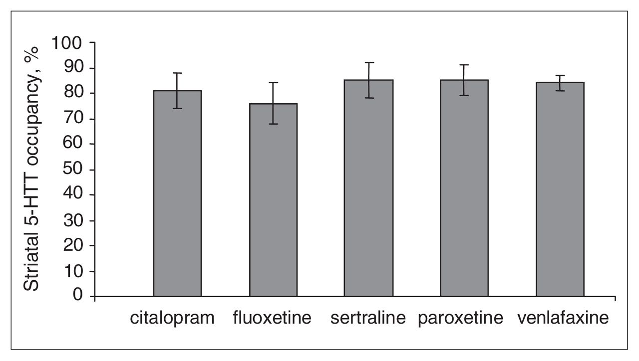
In 2001, the first SSRI occupancy study with [11C]DASB PET found an 80% occupancy in multiple regions after 4 weeks of antidepressant treatment, with doses of paroxetine and citalopram known to have clinical effects that distinguish from placebo.34 Since that time, this result has been replicated in brain regions of reasonable size with fluvoxamine33 as well as fluoxetine, sertraline and venlafaxine35 (see Fig. 4 and Fig. 5). Interestingly, an 80% striatal 5-HTT occupancy occurs at minimum clinical dose, despite the 100-fold range in affinity of the 5 SSRIs for the serotonin transporter.35 Further, the in vitro EC50 does not correlate with affinity.35 This demonstrated that, although affinity is obviously a very important property of a drug, it cannot predict occupancy, even when plasma levels are known.35

5–HTT occupancy at minimum therapeutic dose. Mean striatal serotonin transporter (5-HTT) occupancy for 5 selective serotonin reuptake inhibitors after 4 wk of minimum therapeutic dosing. The vertical ranges represent standard deviation. Subjects received citalopram 20–40 mg (n = 7), fluoxetine 20 mg (n = 4), sertraline 50 mg (n = 3), paroxetine 20 mg (n = 7), venlafaxine XR 75 mg (n = 4). Reprinted from the American Journal of Psychiatry.

Relation between striatal 5-HTT occupancy and dose* or plasma concentration† of citalopram. *†The data were fit using an equation of form f(x) = a*x/(b+x). *The relation between dose and occupancy was highly significant (f(x) = 92*x/(b+x), F1,16 = 127, p < 0.0001). †The relation between plasma level and occupancy was highly significant (f(x) = 96*x/(b+x), F1,16 = 103, p < 0.0001). Reprinted from the American Journal of Psychiatry.
Given the association between the clinically relevant dose and 5-HTT occupancy for all SSRIs, it is now generally believed that an 80% 5-HTT occupancy with an SSRI is therapeutically useful. Consequently, to develop antidepressant drugs with serotonin transporter binding, an 80% serotonin transporter occupancy is considered optimal. This practical approach can be applied to phase I trials to assess whether potential new antidepressant drugs are adequately brain penetrant and to guide dosing selection for subsequent phase II clinical trials.
The 2004 study35 also studied the relation between 5-HTT occupancy and plasma level for 5 commonly prescribed SSRIs. There was increasing occupancy with increasing plasma levels, and occupancy plateaued at the higher doses and higher plasma levels. An important result of this study was that both dose and especially plasma level, had a very strong relation to 5-HTT occupancy. This has several practical clinical applications. First, it is unlikely that inadequate 5-HTT occupancy is a barrier to therapeutic response, because one may simply raise the dose of the SSRI to obtain adequate plasma levels. Second, in clinical circumstances, to estimate 5-HTT occupancy, a 5-HTT imaging study does not need to be completed. Instead, one may use the plasma level and the table in the study to estimate the 5-HTT occupancy of any SSRI.35 Third, given the plateau of 5-HTT occupancy in the clinical dosing range, it is unlikely that 5-HTT occupancy has a strong relation to clinical response within current clinical dosing ranges (as was observed).35
In the [11C]DASB studies of 2001 and 2004, 5-HTT occupancy of SSRIs did not exceed 90%. This raises the question as to whether there is a gap in current therapeutic development, such that SSRIs with extremely high 5-HTT occupancy are not available. Suhara and colleagues reported near 100% 5-HTT occupancy with clomipramine, using [11C](+)McN5652 PET within clinical dosing ranges.33 This has clinical implications because it suggests that clomipramine may be associated with high 5-HTT occupancy within clinical dosing ranges. This result is consistent with the clinical preference for clomipramine or high-dose SSRI for obsessive compulsive disorder191 (for which greater 5-HTT occupancy is preferred).
To date, it seems that 5-HTT occupancy values after short-term dosings largely resemble the findings after 4 weeks of SSRI dosing.33–36,48,169 This has implications for antidepressant development, because it is often desirable to study single - dose occupancy in early phase I investigations, before multiple dosing studies. Results by Parsey and colleagues190 are similar in subcortical regions but may be discrepant in other regions; this may require further study.
Future 5-HTT occupancy investigations are likely to focus on novel antidepressant drugs that bind to both the serotonin transporter and other targets with high affinity.189 An interesting question for future research will be whether an 80% occupancy in reasonably large brain structures is necessary for therapeutic effect when antidepressants target additional therapeutic sites.
Main Findings and Implications
The following are the key findings and/or implications from studies of 5-HTT occupancy:
An 80% regional 5-HTT occupancy, particularly in striatum, typically occurs at doses of SSRIs known to distinguish from placebo in clinical trials of MDD.33–35
Affinity values, even with accompanying blood plasma drug levels, cannot predict 5-HTT occupancy.34,35 5-HTT imaging methods are essential to predict 5-HTT occupancy.
Inadequate 5-HTT occupancy alone cannot adequately explain treatment refractoriness, because 5-HTT occupancy is most strongly related to dose and plasma levels.34,35
5-HTT occupancy will be a useful tool for antidepressant development, either to develop antidepressant drugs for firstline treatment (ideally with near 80% 5-HTT occupancy34,35) or to develop antidepressant drugs with higher 5-HTT occupancy values33 for treatment refractory depression.
Acknowledgements
I would like to thank Alan Wilson who discovered (first to synthesize and characterize) the [11C]DASB compound. I would like to also thank Nathalie Ginovart and Sylvain Houle for their modeling efforts with [11C]DASB. I would also like to thank members of the Toronto PET centre who have actively contributed to studies of serotonin receptors in depression or supporting technology, especially Armando Garcia, Sandra Sagrati, Anahita Carbonneau, Anna Carella, Verdell Goulding, Terry Bell, Ted Harris-Brandts, Alvina Ng and Doug Hussey. Funding support from the Canadian Institutes of Health Research, NARSAD and Eli Lilly Canada was also appreciated.
Footnotes
Medical subject headings: serotonin; serotonin transporter; depression; antidepressant; PET; positron emission tomography
Competing interests: Dr. Meyer has acted as a paid consultant to Lundbeck and GlaxoSmithKline.
- Received August 30, 2006.
- Revision received October 20, 2006.
- Accepted October 22, 2006.
References

Article tools


Related Articles
Cited By...
- No citing articles found.