
Main menu

  • Home
  • Issues
    • Issue in progress
    • Issues by date
  • Sections
    • Editorial
    • Review
    • Research
    • Commentary
    • Psychopharmacology for the Clinician
    • Letters to the Editor
  • Topic Collections
  • Instructions for Authors
    • Overview for authors
    • Submission checklist
    • Editorial policies
    • Publication fees
    • Submit a manuscript
    • Dr. Francis Wayne Quan Memorial Prize
    • Open access
  • Alerts
    • Email alerts
    • RSS
  • About
    • General information
    • Staff
    • Editorial Board
    • Contact
  • CMAJ JOURNALS
    • CMAJ
    • CMAJ Open
    • CJS
    • JAMC

User menu

Search

  • Advanced search
JPN
  • CMAJ JOURNALS
    • CMAJ
    • CMAJ Open
    • CJS
    • JAMC
JPN

Advanced Search

  • Home
  • Issues
    • Issue in progress
    • Issues by date
  • Sections
    • Editorial
    • Review
    • Research
    • Commentary
    • Psychopharmacology for the Clinician
    • Letters to the Editor
  • Topic Collections
  • Instructions for Authors
    • Overview for authors
    • Submission checklist
    • Editorial policies
    • Publication fees
    • Submit a manuscript
    • Dr. Francis Wayne Quan Memorial Prize
    • Open access
  • Alerts
    • Email alerts
    • RSS
  • About
    • General information
    • Staff
    • Editorial Board
    • Contact
  • Subscribe to our alerts
  • RSS feeds
  • Follow JPN on Twitter
Research Paper

Grey matter volume increase following electroconvulsive therapy in patients with late life depression: a longitudinal MRI study

Filip Bouckaert, François-Laurent De Winter, Louise Emsell, Annemieke Dols, Didi Rhebergen, Martien Wampers, Stefan Sunaert, Max Stek, Pascal Sienaert and Mathieu Vandenbulcke
J Psychiatry Neurosci March 01, 2016 41 (2) 105-114; DOI: https://doi.org/10.1503/jpn.140322
Filip Bouckaert
From the Department of Old Age Psychiatry, UPC KU-Leuven, Belgium (Bouckaert, De Winter, Wampers, Vandenbulcke); the GGZ inGeest/Department of Psychiatry and the EMGO+ Institute for Health and Care Research, VU University Medical Center, Amsterdam, The Netherlands (Dols, Rhebergen, Stek); the Department of ECT and Department of Mood Disorders, UPC KULeuven, Belgium (Bouckaert, Sienaert); and Translational MRI, Department of Imaging and Pathology, KU Leuven and Radiology, University Hospitals Leuven, Belgium (Emsell, Sunaert)
  • Find this author on Google Scholar
  • Find this author on PubMed
  • Search for this author on this site
  • For correspondence: [email protected]
François-Laurent De Winter
From the Department of Old Age Psychiatry, UPC KU-Leuven, Belgium (Bouckaert, De Winter, Wampers, Vandenbulcke); the GGZ inGeest/Department of Psychiatry and the EMGO+ Institute for Health and Care Research, VU University Medical Center, Amsterdam, The Netherlands (Dols, Rhebergen, Stek); the Department of ECT and Department of Mood Disorders, UPC KULeuven, Belgium (Bouckaert, Sienaert); and Translational MRI, Department of Imaging and Pathology, KU Leuven and Radiology, University Hospitals Leuven, Belgium (Emsell, Sunaert)
  • Find this author on Google Scholar
  • Find this author on PubMed
  • Search for this author on this site
Louise Emsell
From the Department of Old Age Psychiatry, UPC KU-Leuven, Belgium (Bouckaert, De Winter, Wampers, Vandenbulcke); the GGZ inGeest/Department of Psychiatry and the EMGO+ Institute for Health and Care Research, VU University Medical Center, Amsterdam, The Netherlands (Dols, Rhebergen, Stek); the Department of ECT and Department of Mood Disorders, UPC KULeuven, Belgium (Bouckaert, Sienaert); and Translational MRI, Department of Imaging and Pathology, KU Leuven and Radiology, University Hospitals Leuven, Belgium (Emsell, Sunaert)
  • Find this author on Google Scholar
  • Find this author on PubMed
  • Search for this author on this site
Annemieke Dols
From the Department of Old Age Psychiatry, UPC KU-Leuven, Belgium (Bouckaert, De Winter, Wampers, Vandenbulcke); the GGZ inGeest/Department of Psychiatry and the EMGO+ Institute for Health and Care Research, VU University Medical Center, Amsterdam, The Netherlands (Dols, Rhebergen, Stek); the Department of ECT and Department of Mood Disorders, UPC KULeuven, Belgium (Bouckaert, Sienaert); and Translational MRI, Department of Imaging and Pathology, KU Leuven and Radiology, University Hospitals Leuven, Belgium (Emsell, Sunaert)
  • Find this author on Google Scholar
  • Find this author on PubMed
  • Search for this author on this site
Didi Rhebergen
From the Department of Old Age Psychiatry, UPC KU-Leuven, Belgium (Bouckaert, De Winter, Wampers, Vandenbulcke); the GGZ inGeest/Department of Psychiatry and the EMGO+ Institute for Health and Care Research, VU University Medical Center, Amsterdam, The Netherlands (Dols, Rhebergen, Stek); the Department of ECT and Department of Mood Disorders, UPC KULeuven, Belgium (Bouckaert, Sienaert); and Translational MRI, Department of Imaging and Pathology, KU Leuven and Radiology, University Hospitals Leuven, Belgium (Emsell, Sunaert)
  • Find this author on Google Scholar
  • Find this author on PubMed
  • Search for this author on this site
Martien Wampers
From the Department of Old Age Psychiatry, UPC KU-Leuven, Belgium (Bouckaert, De Winter, Wampers, Vandenbulcke); the GGZ inGeest/Department of Psychiatry and the EMGO+ Institute for Health and Care Research, VU University Medical Center, Amsterdam, The Netherlands (Dols, Rhebergen, Stek); the Department of ECT and Department of Mood Disorders, UPC KULeuven, Belgium (Bouckaert, Sienaert); and Translational MRI, Department of Imaging and Pathology, KU Leuven and Radiology, University Hospitals Leuven, Belgium (Emsell, Sunaert)
  • Find this author on Google Scholar
  • Find this author on PubMed
  • Search for this author on this site
Stefan Sunaert
From the Department of Old Age Psychiatry, UPC KU-Leuven, Belgium (Bouckaert, De Winter, Wampers, Vandenbulcke); the GGZ inGeest/Department of Psychiatry and the EMGO+ Institute for Health and Care Research, VU University Medical Center, Amsterdam, The Netherlands (Dols, Rhebergen, Stek); the Department of ECT and Department of Mood Disorders, UPC KULeuven, Belgium (Bouckaert, Sienaert); and Translational MRI, Department of Imaging and Pathology, KU Leuven and Radiology, University Hospitals Leuven, Belgium (Emsell, Sunaert)
  • Find this author on Google Scholar
  • Find this author on PubMed
  • Search for this author on this site
Max Stek
From the Department of Old Age Psychiatry, UPC KU-Leuven, Belgium (Bouckaert, De Winter, Wampers, Vandenbulcke); the GGZ inGeest/Department of Psychiatry and the EMGO+ Institute for Health and Care Research, VU University Medical Center, Amsterdam, The Netherlands (Dols, Rhebergen, Stek); the Department of ECT and Department of Mood Disorders, UPC KULeuven, Belgium (Bouckaert, Sienaert); and Translational MRI, Department of Imaging and Pathology, KU Leuven and Radiology, University Hospitals Leuven, Belgium (Emsell, Sunaert)
  • Find this author on Google Scholar
  • Find this author on PubMed
  • Search for this author on this site
Pascal Sienaert
From the Department of Old Age Psychiatry, UPC KU-Leuven, Belgium (Bouckaert, De Winter, Wampers, Vandenbulcke); the GGZ inGeest/Department of Psychiatry and the EMGO+ Institute for Health and Care Research, VU University Medical Center, Amsterdam, The Netherlands (Dols, Rhebergen, Stek); the Department of ECT and Department of Mood Disorders, UPC KULeuven, Belgium (Bouckaert, Sienaert); and Translational MRI, Department of Imaging and Pathology, KU Leuven and Radiology, University Hospitals Leuven, Belgium (Emsell, Sunaert)
  • Find this author on Google Scholar
  • Find this author on PubMed
  • Search for this author on this site
Mathieu Vandenbulcke
From the Department of Old Age Psychiatry, UPC KU-Leuven, Belgium (Bouckaert, De Winter, Wampers, Vandenbulcke); the GGZ inGeest/Department of Psychiatry and the EMGO+ Institute for Health and Care Research, VU University Medical Center, Amsterdam, The Netherlands (Dols, Rhebergen, Stek); the Department of ECT and Department of Mood Disorders, UPC KULeuven, Belgium (Bouckaert, Sienaert); and Translational MRI, Department of Imaging and Pathology, KU Leuven and Radiology, University Hospitals Leuven, Belgium (Emsell, Sunaert)
  • Find this author on Google Scholar
  • Find this author on PubMed
  • Search for this author on this site
  • Article
  • Figures & Tables
  • Responses
  • Metrics
  • PDF
Loading

Article Figures & Tables

Figures

  • Tables
  • Fig. 1
    • Download figure
    • Open in new tab
    • Download powerpoint
    Fig. 1

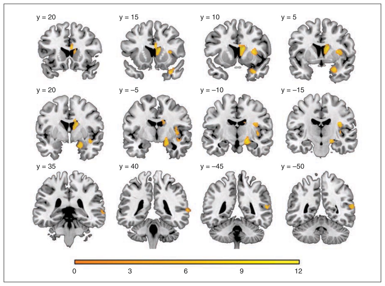
    Grey matter volume change following electroconvulsive therapy: statistical t map threshold at p < 0.05 after family-wise error correction with a cluster extent of 50 voxels displayed on coronal slices. The y coordinate corresponds to Montreal Neurological Institite standard space.

  • Fig. 2
    • Download figure
    • Open in new tab
    • Download powerpoint
    Fig. 2

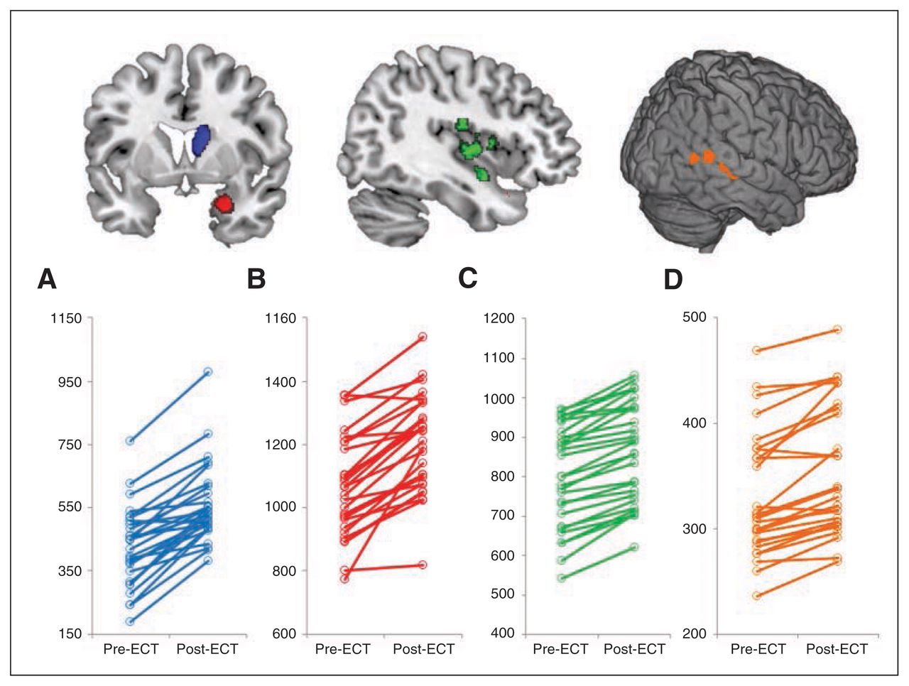
    Total grey matter volume (Y axis in cubic millimetres) within different regions per subset pre- and post–electroconvulsive therapy (ECT) in the (A) caudate cluster, (B) medial temporal cluster, (C) insular clusters and (D) posterior superior temporal clusters.

  • Fig. 3
    • Download figure
    • Open in new tab
    • Download powerpoint
    Fig. 3

    Laterality index (LI) curves for different regions of interest derived from the automated anatomical labels atlas.

  • Fig. 4
    • Download figure
    • Open in new tab
    • Download powerpoint
    Fig. 4

    Correlations between grey matter volume change in the caudate cluster and change in (A) total CORE Assessment score (r = 0.63, p < 0.001), (B) noninteraction subscale score (r = 0.67, p < 0.001), (C) agitation subscale score (r = 0.35, p = 0.06) and (D) retardation subscale score (r = 0.41, p = 0.027).

Tables

  • Figures
    • View popup
    Table 1

    Clinical outcome measures

    Period; mean ± SD (range)
    MeasurePre-ECTPost-ECTChanges; mean ± SD (range)t27p value
    MADRS35.04 ± 6.88 (22–50)9.61 ± 11.47 (0–44)25.43 ± 11.05 (−8 to 45)12.18< 0.001
    MMSE23.64 ± 4.54 (12–30)26.32 ± 3.44 (18–30)−2.68 ± 3.83 (−14 to 6)−3.700.001
    CORE total17.64 ± 8.17 (3–35)4.96 ± 5.00 (0–20)12.68 ± 8.60 (−3 to 31)7.80< 0.001
    CORE cluster noninteraction6.36 ± 4.06 (0–16)1.54 ± 1.87 (0–7)4.82 ± 4.43 (−1 to 16)5.76< 0.001
    CORE cluster agitation3.79 ± 3.14 (0–11)0.89 ± 1.57 (0–7)2.89 ± 3.02 (−1 to 9)5.06< 0.001
    CORE cluster retardation7.5 ± 3.73 (1–17)2.54 ± 2.64 (0–9)4.96 ± 3.30 (−2 to 11)7.95< 0.001
    • ECT = electroconvulsive therapy; MADRS = Montgomery–Åsberg Depression Rating Scale; MMSE = Mini Mental State Examination; SD = standard deviation.

    • View popup
    Table 2

    Grey matter volume changes following ECT

    MNI coordinates
    LocationT*Z*xyzp valueCluster size
    Caudate nucleus12.077.0281116< 0.001941
    Medial temporal lobe11.316.8121−9−14< 0.001769
    Insula (posterior superior)9.966.4038−1321< 0.001170
    Insula (anterior superior)9.286.1733910< 0.001262
    Insula (inferior)7.965.67410−140.00372
    Insula (posterior inferior)7.555.4938−1560.007111
    Post-temporal (middle)8.635.9357−48120.001128
    Post-temporal (posterior)8.525.8957−58130.00154
    Post-temporal (anterior)8.065.7066−3970.00272
    • ECT = electroconvulsive therapy; MNI = Montreal Neurological Institute.

    • ↵* Peak level.

    • View popup
    Table 3

    Correlations between changes in clinical scales and percentage change of grey matter volume

    Region; % change in grey matter volume
    MeasureCaudate nucleusMedial temporal lobeInsulaPosterior temporal region
    Change in MADRSr = 0.26r = 0.19r = 0.18r = 0.17
    p = 0.18p = 0.31p = 0.35p = 0.37
    Change in MMSEr = 0.45r = 0.08r = 0.53r = 0.35
    p = 0.014p = 0.65p = 0.003p = 0.06
    Change in CORE total scorer = 0.63r = 0.15r = 0.47r = 0.03
    p < 0.001*p = 0.44p = 0.010p = 0.88
    Change in CORE noninteraction subscaler = 0.67r = 0.01r = 0.48r = 0.007
    p < 0.001*p = 0.92p = 0.009p = 0.96
    Change in CORE agitation subscaler = 0.35r = 0.40r = 0.29r = 0.06
    p = 0.06p = 0.033p = 0.12p = 0.75
    Change in CORE retardation subscaler = 0.41r = 0.0001r = 0.32r = 0.01
    p = 0.027p = 0.99p = 0.09p = 0.95
    No. of ECT sessionsr = 0.18r = 0.29r = 0.33r = 0.09
    p = 0.35p = 0.13p = 0.07p = 0.62
    • ECT = electroconvulsive therapy; MADRS = Montgomery–Åsberg Depression Rating Scale; MMSE = Mini Mental State Examination.

    • ↵* Significant after Bonferroni correction (adjusted p = 0.001).

PreviousNext
Back to top

In this issue

Journal of Psychiatry and Neuroscience: 41 (2)
J Psychiatry Neurosci
Vol. 41, Issue 2
1 Mar 2016
  • Table of Contents
  • Index by author

Article tools

Respond to this article
Print
Download PDF
Article Alerts
To sign up for email alerts or to access your current email alerts, enter your email address below:
Email Article

Thank you for your interest in spreading the word on JPN.

NOTE: We only request your email address so that the person you are recommending the page to knows that you wanted them to see it, and that it is not junk mail. We do not capture any email address.

Enter multiple addresses on separate lines or separate them with commas.
Grey matter volume increase following electroconvulsive therapy in patients with late life depression: a longitudinal MRI study
(Your Name) has sent you a message from JPN
(Your Name) thought you would like to see the JPN web site.
CAPTCHA
This question is for testing whether or not you are a human visitor and to prevent automated spam submissions.
Citation Tools
Grey matter volume increase following electroconvulsive therapy in patients with late life depression: a longitudinal MRI study
Filip Bouckaert, François-Laurent De Winter, Louise Emsell, Annemieke Dols, Didi Rhebergen, Martien Wampers, Stefan Sunaert, Max Stek, Pascal Sienaert, Mathieu Vandenbulcke
J Psychiatry Neurosci Mar 2016, 41 (2) 105-114; DOI: 10.1503/jpn.140322

Citation Manager Formats

  • BibTeX
  • Bookends
  • EasyBib
  • EndNote (tagged)
  • EndNote 8 (xml)
  • Medlars
  • Mendeley
  • Papers
  • RefWorks Tagged
  • Ref Manager
  • RIS
  • Zotero
‍ Request Permissions
Share
Grey matter volume increase following electroconvulsive therapy in patients with late life depression: a longitudinal MRI study
Filip Bouckaert, François-Laurent De Winter, Louise Emsell, Annemieke Dols, Didi Rhebergen, Martien Wampers, Stefan Sunaert, Max Stek, Pascal Sienaert, Mathieu Vandenbulcke
J Psychiatry Neurosci Mar 2016, 41 (2) 105-114; DOI: 10.1503/jpn.140322
Digg logo Reddit logo Twitter logo Facebook logo Google logo Mendeley logo
  • Tweet Widget
  • Facebook Like

Related Articles

  • No related articles found.
  • PubMed
  • Google Scholar

Cited By...

  • Biophysical mechanisms of electroconvulsive therapy-induced volume expansion in the medial temporal lobe: a longitudinal in vivo human imaging study
  • Electroconvulsive Therapy and Brain Damage: Survey of the Evidence From a Philosophical Promontory
  • Google Scholar

Similar Articles

Content

  • Current issue
  • Past issues
  • Collections
  • Alerts
  • RSS

Authors & Reviewers

  • Overview for Authors
  • Submit a manuscript
  • Manuscript Submission Checklist

About

  • General Information
  • Staff
  • Editorial Board
  • Contact Us
  • Advertising
  • Reprints
  • Copyright and Permissions
  • Accessibility
  • CMA Civility Standards
CMAJ Group

Copyright 2023, CMA Impact Inc. or its licensors. All rights reserved. ISSN 1180-4882.

All editorial matter in JPN represents the opinions of the authors and not necessarily those of the Canadian Medical Association or its subsidiaries.
To receive any of these resources in an accessible format, please contact us at CMAJ Group, 500-1410 Blair Towers Place, Ottawa ON, K1J 9B9; p: 1-888-855-2555; e: [email protected].
View CMA's Accessibility policy.

Powered by HighWire