

Article Figures & Tables
Figures
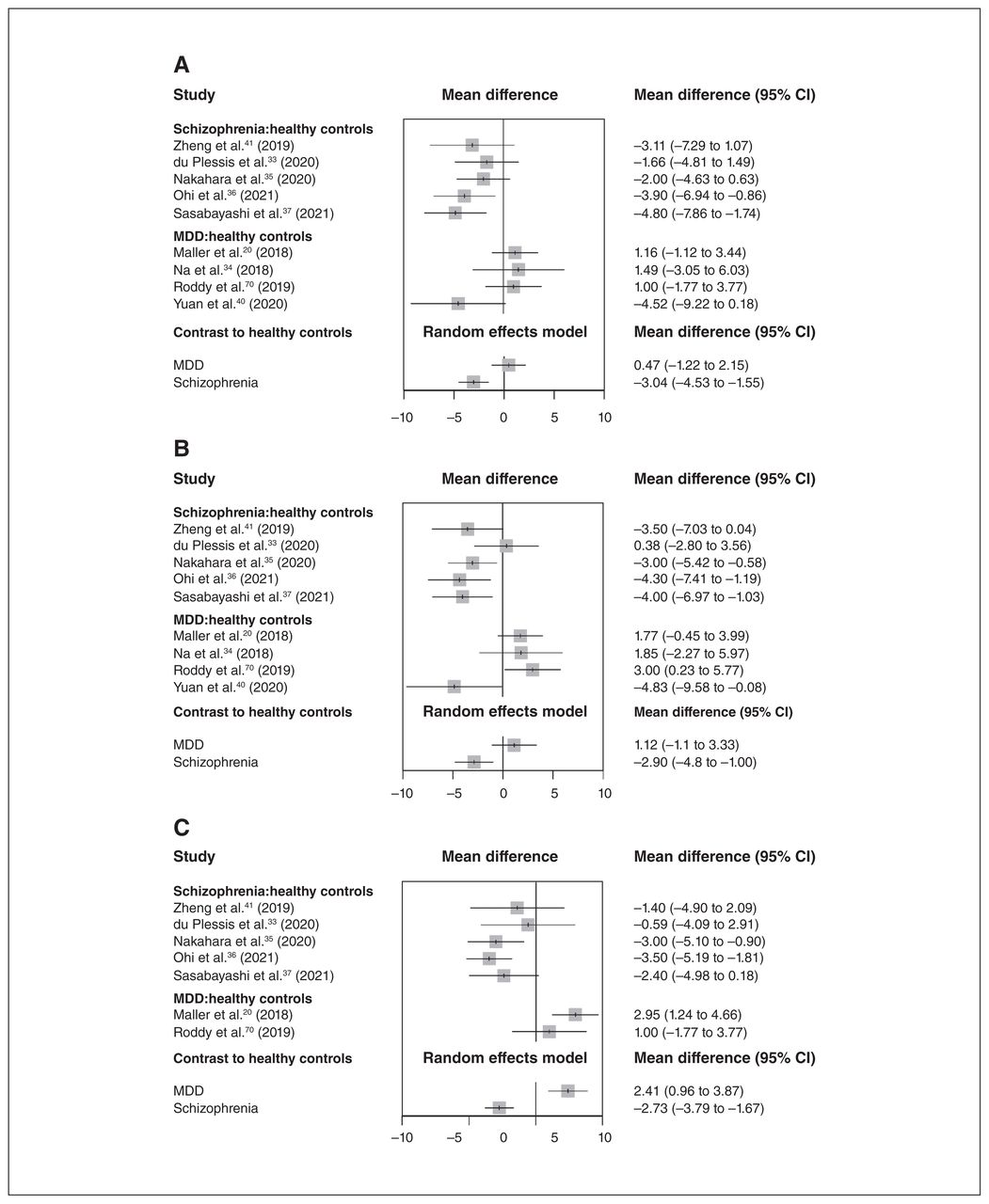
- Figure 1
(A) Forest plot for the left parasubiculum. (B) Forest plot for the right parasubiculum. (C) Forest plot for the right HATA. CI = confidence interval; HATA = hippocampus–amygdala transition area; MDD = major depressive disorder.
Tables
Author (year) Participants, n (% female, mean age) MRI scanner Segmentation method Study design Main findings Alnæs et al. (42) (2019)*† Schizophrenia, 1151 (31%, 34 y)
Healthy controls, 2010 (44%, 33 y)1.5 T and 3.0 T Automatic (FreeSurfer 6.0¶) Cross-sectional design, case–control, cohort, multisite; genes Smaller volume in all subfields; larger left and right hippocampal fissure Brown et al. (43) (2019) MDD, 24 (38%, 40 y)
Healthy controls, 20 (25%, 40 y)7.0 T Automatic (FreeSurfer 6.0¶) Cross-sectional design, case–control, cohort No significant difference Cao et al. (44) (2018) MDD, 24 (58%, 31 y)
Healthy controls, 15 (66%, 33 y)3.0 T Automatic (FreeSurfer 5.3¶) Longitudinal design, case–control; ECT Baseline: no significant difference Longitudinal changes: increased left CA2/3, left and right CA4, left and right granule cell layer of the dentate gyrus, left subiculum Cao et al. (31) (2017) MDD, 86 (70%, 41 y)
Healthy controls, 152 (63%, 35 y)1.5 T Automatic (FreeSurfer 5.3¶) Cross-sectional design, case–control No significant difference Doolin et al. (32) (2018) MDD, 74 (64%, 33 y)
Healthy controls, 37 (51%, 31 y)3.0 T Automatic (FreeSurfer 6.0¶) Cross-sectional design, case–control, cohort Smaller left CA1, left and right CA2/3, right CA4 du Plessis et al. (33) (2020) First-episode schizophrenia, 79 (27%, 23 y)
Healthy controls, 82 (43 %, 23)3.0 T Automatic (FreeSurfer 6.0¶) Cross-sectional design, case–control, cohort Statistical analysis between 2 groups was not available Frodl et al. (45) (2014) MDD, 43 (60%, 41 y)
Healthy controls, 43 (60%, 37 y)3.0 T Automatic (FreeSurfer**) Cross-sectional design, case–control; genes Smaller CA1, CA2/3, CA4/dentate gyrus, subiculum Frodl et al. (46) (2014) MDD, 38 (66%, 41 y)
Healthy controls, 44 (61%, 36 y)3.0 T Automatic (FreeSurfer**) Cross-sectional design, case–control; genes Smaller left CA2/3, left CA4/dentate gyrus Han et al. (21) (2019)‡ MDD, 102 (59%, 36 y)
Healthy controls, 135 (58%, 36 y)3.0 T Automatic (FreeSurfer 6.0¶) Cross-sectional design, case–control Smaller left and right whole hippocampus, left and right CA1, left CA2/3, left and right CA4, left and right granule cell layer of the dentate gyrus, right subiculum, right presubiculum, left and right molecular layer Han et al. (47) (2017)‡ MDD, 105 (82%, 43 y)
Healthy controls, 85 (71%, 40 y)3.0 T Automatic (FreeSurfer 5.3¶) Cross-sectional design, case–control; genes No significant difference Han et al. (48) (2016) MDD, 20 (100%, 42 y)
Healthy controls, 21 (100%, 42 y)1.5 T Automatic (FreeSurfer 5.3**) Cross-sectional design, case–control Smaller left whole hippocampus, left CA2/3, left CA4/dentate gyrus, left and right subiculum Harel et al. (49) (2016) MDD, 15 (53%, 36 y)
Healthy controls, 15 (47%, 37 y)3.0 T Automatic (FreeSurfer 5.3**) Cross-sectional design, case–control Smaller right whole hippocampus, right CA1, right CA2/3, right CA4/dentate gyrus Ho et al. (16) (2017) Schizophrenia, 155 (32%, 32 y)
Healthy controls, 79 (35%, 31 y)3.0 T Automatic (FreeSurfer 5.3¶) Cross-sectional design, case–control; longitudinal design in a subcohort; multisite Schizophrenia: smaller left and right whole hippocampus, left CA1 Early-course schizophrenia: smaller left and right whole hippocampus, left and right CA1, right granule cell layer of the dentate gyrus
Longitudinal changes: decreased left and right CA1, right CA2/3, left and right granule cell layer of the dentate gyrus, right molecular layerSchizophrenia, 46 (22%, 43 y)
Healthy controls, 46 (22%, 42 y)Smaller left and right whole hippocampus, left and right CA1, left and right CA2/3, left and right CA4, left and right granule cell layer of the dentate gyrus, left and right subiculum, left and right molecular layer, left and right hippocampal tail Hu et al. (66) (2020) Never-treated long-term schizophrenia, 29 (55%, 46 y)
Treated long-term schizophrenia, 40 (55%, 48 y)
Healthy controls, 40 (55%, 48 y)3.0 T Automatic (FreeSurfer 6.0¶) Cross-sectional design, case–control Never-treated long-term schizophrenia: smaller left and right whole hippocampus, right CA1 (body), left and right CA2/3 (head), right CA2/3 (body), left and right CA4 (head), left and right CA4 (body), left and right granule cell layer of the dentate gyrus (head), left and right granule cell layer of the dentate gyrus (body), left and right subiculum (body), left and right molecular layer (body), left and right hippocampal tail
Treated long-term schizophrenia: smaller left and right whole hippocampus, left CA4 (body), left granule cell layer of the dentate gyrus (body), left and right subiculum (body), left and right molecular layer (body), left and right hippocampal tailHu et al. (50) (2019) Nonresponding MDD, 13 (38%, 36 y)
Early responding MDD, 25 (44%, 36 y)
Healthy controls, 55 (62%, 36 y)3.0 T Automatic (FreeSurfer 5.3**) Cross-sectional design, case–control, cohort MDD: no significant difference Nonresponding MDD: larger left and right CA1, left CA2/3, left CA4/dentate gyrus, left and right subiculum Huang et al. (67) (2013)§ Unmedicated MDD, 9 (44%, 33 y)
Medicated MDD, 11 (55%, 37 y)
Healthy controls, 27 (70%, 33 y)4.7 T Manual Cross-sectional design, case–control Unmedicated MDD: smaller CA1–3 (body), dentate gyrus Hýža et al. (51) (2016) First-episode schizophrenia, 58 (0%, 23 y)
Healthy controls, 58 (0%, 24 y)1.5 T Automatic (FreeSurfer 5.2**) Cross-sectional design, case–control Larger left CA1 Jiang et al. (52) (2019) Schizophrenia with symptom remission after ECT, 10 (50%, 30 y)
Schizophrenia without symptom remission after ECT, 11 (55%, 28 y)
Schizophrenia treated by ECT (with and without symptom remission), 21 (52%, 29 y)
Schizophrenia with symptom remission after antipsychotic medication, 12 (75%, 31 y)
Schizophrenia without symptom remission after antipsychotic medication, 9 (33%, 30 y)
Schizophrenia treated by antipsychotic medication (with and without symptom remission), 21 (57%, 31 y)
Healthy controls, 23 (52%, 31 y)3.0 T Automatic (FreeSurfer 6.0¶) Longitudinal design, cohort; ECT Longitudinal changes: schizophrenia treated by ECT (with and without symptom remission), increased left and right whole hippocampus Kakeda et al. (53) (2018) First-episode MDD, 40 (50%, 47 y)
Healthy controls, 47 (28%, 41 y)3.0 T Automatic (FreeSurfer 6.0¶) Cross-sectional design, case–control No significant difference Kawano et al. (68) (2015) First-episode schizophrenia, 19 (53%, 25 y)
Subchronic schizophrenia, 6 (50%, 22 y)
Chronic schizophrenia, 9 (33%, 37 y)
Healthy controls, 15 (33%, 25 y)1.5 T Automatic (FreeSurfer 5.1**) Cross-sectional design, case–control; longitudinal design in a subcohort Baseline: first-episode schizophrenia, smaller left CA2/3, left CA4/ dentate gyrus
Subchronic schizophrenia: smaller left whole hippocampus, left CA2/3, left CA4/ dentate gyrus
Chronic schizophrenia: smaller left whole hippocampus, left CA2/3, left CA4/ dentate gyrusKraus et al. (54) (2019) Acute MDD, 20 (70%, 31 y)
Remitted MDD, 28 (57%, 27 y)
Healthy controls, 22 (55%, 26 y)7 T Automatic (FreeSurfer 6.0¶) Longitudinal design, open-label trial Time 1: remitted MDD, larger right hippocampal fissure
Longitudinal changes: no significant difference
Time 2: remitted MDD, larger right HATALi et al. (55) (2018) First-episode schizophrenia, 41 (59%, 24 y)
Healthy controls, 39 (51%, 24 y)3.0 T Automatic (FreeSurfer 6.0¶) Cross-sectional design, case–control; longitudinal design; antipsychotics Baseline: larger left and right CA4, left and right granule cell layer of the dentate gyrus, left and right molecular layer
Longitudinal changes: decreased left and right whole hippocampus, left CA1, left CA2/3, left and right CA4, left and right granule cell layer of the dentate gyrus, left and right molecular layer, left and right hippocampal tail, left fimbria
After treatment: larger left and right CA4Maller et al. (20) (2018) MDD, 182 (52%, 33 y)
Healthy controls, 68 (50%, 30 y)3.0 T Automatic (FreeSurfer 6.0¶) Open-label trial Baseline: larger hippocampal tail Mathew et al. (69) (2014) Schizophrenia, 219 (34%, 35 y)
Healthy controls, 337 (55%, 37 y)Unknown Automatic (FreeSurfer 5.1**) Cross-sectional design, case–control, multisite Smaller left and right whole hippocampus, left and right CA1, left and right CA2/3, left and right CA4/dentate gyrus, left and right subiculum, left and right presubiculum Mikolas et al. (56) (2019) MDD, 85 (67%, 39 y)
Healthy controls, 67 (67%, 36 y)3.0 T Automatic (FreeSurfer 5.3¶) Cross-sectional design, case–control; genes Smaller whole hippocampus, CA1, CA2/3, CA4, granule cell layer of the dentate gyrus, molecular layer Na et al. (57) (2014) MDD, 45 (76%, 42 y)
Healthy controls, 72 (71%, 41 y)3.0 T Automatic (FreeSurfer 5.0**) Cross-sectional design, case–control; genes No significant difference Na et al. (34) (2018) MDD, 47 (100%, 45 y)
Healthy controls, 30 (100%, 43 y)3.0 T Automatic (FreeSurfer 6.0¶) Cross-sectional design, case–control; genes No significant difference Nakahara et al. (35) (2020) Schizophrenia, 176 (25%, 39 y)
Healthy controls, 173 (29%, 38 y)3.0 T Automatic (FreeSurfer 6.0¶) Cross-sectional design, case–control, multisite Smaller CA1, CA4, granule cell layer of the dentate gyrus, molecular layer, hippocampal tail; larger hippocampal fissure Nguyen et al. (58) (2019) First-episode MDD, 38 (47%, 47 y)
Healthy controls, 39 (28%, 41 y)3.0 T Automatic (FreeSurfer 6.0¶) Cross-sectional design, case–control; genes No significant difference Ohi et al. (36) (2021) Schizophrenia, 138 (60%, 42 y)
Healthy controls, 162 (33%, 37 y)3.0 T Automatic (FreeSurfer 6.0¶) Cross-sectional design, case–control Smaller right HATA; larger right hippocampal fissure Orfei et al. (59) (2017) Schizophrenia, 45 (33%, 40 y)
Healthy controls, 45 (33%, 40 y)3.0 T Automatic (FreeSurfer**) Cross-sectional design, case–control Smaller left and right whole hippocampus, left and right CA1, left and right CA2/3, left and right CA4/dentate gyrus, left subiculum, left and right presubiculum, left and right hippocampal fissure Ota et al. (23) (2017) Schizophrenia, 20 (25%, 37 y)
MDD, 36 (47%, 38 y)
Healthy controls, 35 (46%, 39 y)3.0 T Automatic (ASHS) Cross-sectional design, case–control Schizophrenia: smaller whole hippocampus, CA1, dentate gyrus than healthy controls, smaller whole hippocampus, dentate gyrus than MDD without medication MDD: no significant difference Otsuka et al. (60) (2019) First-episode MDD, 27 (41%, 46 y);
Healthy controls, 42 (26%, 41 y)3.0 T Automatic (FreeSurfer 6.0¶) Cross-sectional design, case–control; genes No significant difference Roddy et al. (70) (2019) MDD, 80 (71%, 35 y)
Healthy controls, 83 (59%, 32 y)3.0 T Automatic (FreeSurfer 6.0¶) Cross-sectional design, case–control Smaller left CA1, left and right CA2/3, left and right CA4, left and right granule cell layer of the dentate gyrus, left and right subiculum, left hippocampal tail; larger right molecular layer Sasabayashi et al. (37) (2021) Schizophrenia, 77 (49%, 29 y)
Healthy controls, 87 (47%, 26 y)3.0 T Automatic (FreeSurfer 6.0¶) Cross-sectional design, case–control, cohort Smaller left and right CA1, left and right molecular layer, left hippocampal tail Tannous et al. (71) (2020) MDD, 71 (55%, 32 y)
Healthy controls, 46 (54%, 32 y)7.0 T Automatic (FreeSurfer 6.0¶) Cross-sectional design, case–control, cohort No significant difference Tesli et al. (61) (2020)† Schizophrenia with a history of violence, 24 (4%, 34 y)
Schizophrenia with no history of violence, 51 (2%, 29 y)
Healthy controls, 90 (3%, 33 y)3.0 T Automatic (FreeSurfer 6.0¶) Cross-sectional design, case–control, cohort, multisite Schizophrenia with a history of violence: smaller whole hippocampus, CA1, molecular layer, fimbria, HATA; larger hippocampal fissure
Schizophrenia with no history of violence: no significant differenceTravis et al. (62) (2015)§ MDD, 15 (80%, 38 y)
Healthy controls, 15 (67%, 35 y)4.7 T Manual Cross-sectional design, case–control, cohort Smaller dentate gyrus (body) Travis et al. (63) (2016)§ MDD, 14 (64%, 36 y)
Healthy controls, 14 (71%, 33 y)4.7 T Manual Cross-sectional design, case–control, cohort No significant difference Vargas et al. (64) (2018) Schizophrenia, 91 (26%, 38 y)
Healthy controls, 70 (56%, 18 y)3.0 T Automatic (FreeSurfer 5.3**) Cross-sectional design, case–control Smaller left and right whole hippocampus, right CA1, left and right CA2/3, left and right CA4/dentate gyrus, left and right subiculum, left and right presubiculum Xiu et al. (38) (2021) First-episode schizophrenia, 39 (59%, 29 y)
Healthy controls, 30 (57%, 28 y)3.0 T Automatic (FreeSurfer 5.3¶) Cross-sectional design, case–control No significant difference Xu et al. (39) (2018) MDD, 15 (100%, 35 y)
Healthy controls, 12 (100%, 34 y)3.0 T Automatic (FreeSurfer 6.0¶) Cross-sectional design, case–control Smaller left fimbria Yuan et al. (40) (2020) MDD, 41 (59%, 35 y)
Healthy controls, 44 (59%, 33 y)3.0 T Automatic (FreeSurfer 6.0¶) Cross-sectional design, case–control No significant difference Zheng et al. (41) (2019)* Schizophrenia, 69 (16%, 38 y)
Healthy controls, 72 (29%, 36 y)3.0 T Automatic (FreeSurfer 6.0¶) Cross-sectional design, case–control Smaller left and right whole hippocampus, left CA1, left subiculum, left and right presubiculum, right parasubiculum, left and right molecular layer, left hippocampal tail Zhou et al. (65) (2020) MDD, 44 (64%, 35 y) Healthy controls, 45 (53%, 33 y) 3.0 T Automatic (FreeSurfer 6.0¶) Longitudinal design; ketamine treatment Baseline: smaller left and right whole hippocampus Longitudinal changes: increased right whole hippocampus, right CA4 (head), left CA4 (body), left granule cell layer of the dentate gyrus (body), right molecular layer (head) ASHS = Automatic Segmentation of Hippocampal Subfields; CA = cornu ammonis; ECT = electroconvulsive therapy; HATA = hippocampus–amygdala transition area; MDD = major depressive disorder.
↵* Overlapped data.
↵† Overlapped data.
↵‡ Overlapped data.
↵§ Overlapped data.
↵** Atlas by van Leemput and colleagues. (72)
↵¶ Atlas by Iglesias and colleagues. (15)
- Table 2:
Main characteristics of the studies included in the network meta-analysis (part 1 of 2)
Author (year) No. participants (female) Participant age, y Age at onset, y Illness duration, y Mean no. of episodes Score of severity (scale type) Medication status Substance misuse MRI field strength, subfield segmentation Study design Patients Healthy controls p for sex Patients Healthy controls p for age Ho et al. (16) (2017) 155 (49) 79 (28) 0.66 32.5 31.2 0.30 25.9 6.6 NA 40.6 (PANSS) CPZ-eq: 212.32 ± 191.25 mg/d No substance misuse 3 mo preceding the study 3.0 T, automatic (FreeSurfer 5.3) Cross-sectional design; case–control; longitudinal in a subcohort; multisite 46 (10) 46 (10) > 0.99 42.9 41.9 0.61 24.5 18.2 NA 81.6 (PANSS) CPZ-eq: 532.45 ± 447.16 mg/d Zheng et al. (41) (2019) 69 (11) 72 (21) 0.06 37.7 35.9 0.046 NA NA NA NA NA NA 3.0 T, automatic (FreeSurfer 6.0) Cross-sectional design; case–control du Plessis et al. (33) (2020) 79 (21) 82 (35) 0.03 23.0 23.0 0.97 22.36* 0.64 First-episode schizophrenia 91.27 (PANSS) Treated ≤ 1 mo First-episode schizophrenia: 35 (44%)
Controls: 22 (27%)3.0 T, automatic (FreeSurfer 6.0) Cross-sectional design; case–control; cohort Nakahara et al. (35) (2020) 176 (44) 173 (50) 0.39 38.9 37.6 0.27 21.9 17.1 NA 57.9 (PANSS) CPZ-eq: 372 ± 390 mg/d (n = 144) None 3.0 T, automatic (FreeSurfer 6.0) Cross-sectional design; case–control; multisite Ohi et al. (36) (2021) 138 (83) 162 (54) < 0.05† 42.0 36.7 < 0.05† 26.2 16.3 NA 33.9 (PANSS positive symptoms and PANSS negative symptoms) CPZ-eq: 519.0 ± 524.0 mg/d NA 3.0 T, automatic (FreeSurfer 6.0) Cross-sectional design; case–control Sasabayashi et al. (37) (2021) 77 (38) 87 (41) > 0.05† 28.8 26.3 < 0.05† 22.8 5.6 NA 68.7 (PANSS) HPD-eq: 10.6 ± 8.3 mg/d (n = 65) None 3.0 T, automatic (FreeSurfer 6.0) Cross-sectional design; case–control; cohort Xiu et al. (38) (2021) 39 (23) 30 (17) 0.85 28.9 27.5 0.54 26.95* 1.95 First-episode schizophrenia 44.5 (MCCB) All naive None 3.0 T, automatic (FreeSurfer 5.3) Cross-sectional design; case–control Cao et al. (31) (2017) 86 (60) 152 (96) > 0.05† 41.2 35.4 < 0.05† 32.3* 8.9 3‡ 10.5 (HDRS-17) On medication: 4 Drug use disorder < 10 1.5 T, automatic (FreeSurfer 5.3) Cross-sectional design; case–control Doolin et al. (32) (2018) 74 (47) 37 (19) > 0.05 32.9 30.9 > 0.05 NA NA First-episode MDD (n = 39) > 1 (n = 35) 23.46 (HDRS-17) On medication: 47 Medication-free: 27 NA 3.0 T, automatic (FreeSurfer 6.0) Cross-sectional design; case–control; cohort; Maller et al. (20) (2018) 182 (95) 68 (34) > 0.05 33.0 29.6 0.048 22.2* 10.8 9.6 21.36 (HDRS-17) All naive or with a washout period ≥ 5 half-lives NA 3.0 T, automatic (FreeSurfer 6.0) Open-label trial Na et al. (34) (2018) 47 (47) 30 (30) > 0.05 45.3 43.0 0.41 40.6* 4.7 NA 12.9 (HDRS-17) On medication: 35 NA 3.0 T, automatic (FreeSurfer 6.0) Cross-sectional design; case–control; genes Xu et al. (39) (2018) 15 (15) 12 (12) > 0.05 34.6 34.1 0.51 NA NA NA 33.13 (HDRS-24) NA None 3.0 T, automatic (FreeSurfer 6.0) Cross-sectional design; case–control Han et al. (21) (2019) 102 (60) 135 (78) > 0.05† 36.0 36.0 > 0.05† 32.3* 3.7 First-episode MDD (n = 25) > 1 (n = 77) 13.93 (HDRS-17) On medication: most None 3.0 T, automatic (FreeSurfer 6.0) Cross-sectional design; case–control Roddy et al. (70) (2019) 80 (57) 83 (49) 0.14 34.5 31.5 0.13 32.1* 2.4 First-episode MDD (n = 43) > 1 (n = 37) 22.2 (HDRS-17) NA None 3.0 T, automatic (FreeSurfer 6.0) Cross-sectional design; case–control Yuan et al. (40) (2020) 41 (24) 44 (26) 0.96 34.8 33.3 0.54 18.3* 16.5§ < 3 (n = 18), ≥ 3 (n = 13)¶ 18.3 (HDRS-17) Naive: 16 Untreated ≥ 3 w: 25 MDD with drug use disorder: 17 MDD without drug use disorder: 24 3.0 T, automatic (FreeSurfer 6.0) Cross-sectional design; case–control CPZ-eq = chlorpromazine equivalent; HDRS-17 or -24 = 17- or 24-item Hamilton Depression Rating Scale; HPD-eq = haloperidol equivalent; MCCB = MATRICS Consensus Cognitive Battery; MDD = major depressive disorder; NA = not available; PANSS = Positive and Negative Syndrome Scale.
↵* Determined based on the difference between patient mean age and illness duration.
↵† Inferred based on the results of the corresponding analysis of variance or χ2 test.
↵‡ Median.
↵§ One participant lacked information on illness duration.
↵¶ Ten participants lacked information on number of prior episodes.
- Table 3
Direct volume comparisons between patients with schizophrenia and healthy controls
Region of interest Mean difference (95% CI), mm3 p value* I2, % No. of studies No. of patients with schizophrenia No. of healthy controls Left whole hippocampus† −127.840 (−209.381 to −46.298) 0.005 49 6 700 649 Left CA1† −21.721 (−36.328 to −7.113) 0.006 0 7 779 731 Left CA3 −4.555 (−9.580 to 0.471) 0.08 42 7 779 731 Left CA4† −6.875 (−11.728 to −2.021) 0.008 45 7 779 731 Left granule cell layer of the dentate gyrus† −8.430 (−14.089 to −2.771) 0.006 44 7 779 731 Left subiculum† −10.245 (−18.124 to −2.366) 0.01 36 7 779 731 Left presubiculum† −7.667 (−14.219 to −1.116) 0.03 2 6 578 606 Left parasubiculum† −3.041 (−4.531 to −1.551) < 0.001 0 5 539 576 Left molecular layer† −17.412 (−27.110 to −7.714) 0.002 0 7 779 731 Left hippocampal tail† −24.456 (−40.346 to −8.566) 0.006 74 7 779 731 Left fimbria −2.008 (−5.401 to 1.385) 0.25 20 5 539 576 Left hippocampal fissure† 4.941 (1.953 to 7.930) 0.004 0 5 539 576 Left HATA† −2.044 (−3.156 to −0.932) 0.002 23 5 539 576 Right whole hippocampus† −108.803 (−182.105 to −35.500) 0.01 35 6 700 649 Right CA1† −16.337 (−29.533 to −3.140) 0.02 32 7 779 731 Right CA3 −2.776 (−7.850 to 2.299) 0.28 30 7 779 731 Right CA4 −4.929 (−10.152 to 0.293) 0.07 47 7 779 731 Right granule cell layer of the dentate gyrus† −6.657 (−11.976 to −1.338) 0.02 38 7 779 731 Right subiculum† −8.764 (−17.057 to −0.472) 0.049 8 7 779 731 Right presubiculum −7.051 (−14.077 to −0.024) 0.06 0 6 578 606 Right parasubiculum† −2.899 (−4.797 to −1.001) 0.01 27 5 539 576 Right molecular layer† −13.845 (−25.233 to −2.457) 0.02 23 7 779 731 Right hippocampal tail† −17.221 (−31.208 to −3.234) 0.02 18 7 779 731 Right fimbria† −4.201 (−6.586 to −1.816) 0.003 0 5 539 576 Right hippocampal fissure† 9.849 (6.728 to 12.970) < 0.001 0 5 539 576 Right HATA† −2.727 (−3.787 to −1.667) < 0.001 0 5 539 576 Region of interest Mean difference (95% CI), mm3 p value* I2, % No. of studies No. of patients with MDD No. of healthy controls Left whole hippocampus −74.998 (−175.651 to 25.656) 0.26 88 5 422 325 Left CA1 −13.373 (−28.894 to 2.147) 0.24 83 8 598 546 Left CA3† −7.491 (−12.873 to −2.109) 0.04 71 8 598 544 Left CA4† −8.137 (−13.724 to −2.549) 0.04 69 7 557 504 Left granule cell layer of the dentate gyrus −6.711 (−13.427 to 0.005) 0.22 74 6 535 508 Left subiculum −7.580 (−16.215 to 1.055) 0.24 66 8 596 546 Left presubiculum 0.247 (−6.656 to 7.150) 0.94 72 5 494 466 Left parasubiculum 0.465 (−1.220 to 2.151) 0.77 40 4 347 222 Left molecular layer −2.925 (−15.559 to 9.710) 0.77 81 5 493 464 Left hippocampal tail −3.943 (−25.319 to 17.433) 0.78 88 4 445 436 Left fimbria −4.641 (−10.825 to 1.543) 0.26 74 2 91 92 Left hippocampal fissure 4.000 (−1.544 to 9.544) 0.26 NA 1 75 79 Left HATA 0.628 (−1.139 to 2.396) 0.70 0 2 258 149 Right whole hippocampus −39.031 (−127.489 to 49.426) 0.50 84 5 421 325 Right CA1 −9.269 (−22.842 to 4.303) 0.29 71 8 598 547 Right CA3 −4.766 (−10.052 to 0.520) 0.17 66 8 598 548 Right CA4 −7.333 (−13.175 to −1.491) 0.09 68 7 557 502 Right granule cell layer of the dentate gyrus −6.295 (−12.299 to −0.292) 0.17 62 6 535 508 Right subiculum −8.339 (−17.172 to 0.495) 0.17 77 8 597 547 Right presubiculum 2.210 (−4.963 to 9.384) 0.65 80 5 495 466 Right parasubiculum 1.117 (−1.101 to 3.335) 0.47 63 4 348 223 Right molecular layer 1.735 (−12.626 to 16.095) 0.81 84 5 495 466 Right hippocampal tail 2.729 (−15.585 to 21.042) 0.81 89 4 448 436 Right fimbria −4.211 (−9.167 to 0.745) 0.18 0 2 92 92 Right hippocampal fissure 5.000 (−0.544 to 10.544) 0.17 NA 1 78 81 Right HATA† 2.413 (0.958 to 3.868) 0.01 27 2 260 149 - Table 5
Indirect volume comparisons between patients with schizophrenia and patients with MDD
Region of interest Mean difference (95% CI), mm3 p value* Left whole hippocampus −52.842 (−182.380 to 76.696) 0.67 Left CA1 −8.347 (−29.661 to 12.966) 0.67 Left CA3 2.936 (−4.428 to 10.300) 0.67 Left CA4 1.262 (−6.139 to 8.663) 0.77 Left granule cell layer of the dentate gyrus −1.719 (−10.501 to 7.063) 0.77 Left subiculum −2.665 (−14.354 to 9.025) 0.77 Left presubiculum −7.914 (−17.431 to 1.602) 0.34 Left parasubiculum† −3.506 (−5.755 to −1.256) 0.03 Left molecular layer −14.488 (−30.416 to 1.440) 0.32 Left hippocampal tail −20.513 (−47.148 to 6.122) 0.34 Left fimbria 2.633 (−4.420 to 9.687) 0.67 Left hippocampal fissure 0.941 (−5.357 to 7.239) 0.77 Left HATA −2.672 (−4.761 to −0.584) 0.08 Right whole hippocampus −69.772 (−184.654 to 45.111) 0.43 Right CA1 −7.067 (−25.997 to 11.863) 0.75 Right CA3 1.990 (−5.337 to 9.317) 0.77 Right CA4 2.404 (−5.432 to 10.240) 0.77 Right granule cell layer of the dentate gyrus −0.362 (−8.383 to 7.659) 1.00 Right subiculum −0.426 (−12.542 to 11.691) 1.00 Right presubiculum −9.261 (−19.302 to 0.780) 0.25 Right parasubiculum† −4.016 (−6.935 to −1.097) 0.046 Right molecular layer −15.580 (−33.907 to 2.748) 0.25 Right hippocampal tail −19.950 (−42.993 to 3.094) 0.25 Right fimbria 0.011 (−5.489 to 5.510) 1.00 Right hippocampal fissure 4.849 (−1.513 to 11.211) 0.29 Right HATA† −5.140 (−6.940 to −3.340) < 0.001
In this issue

Article tools


Related Articles
Cited By...
- No citing articles found.